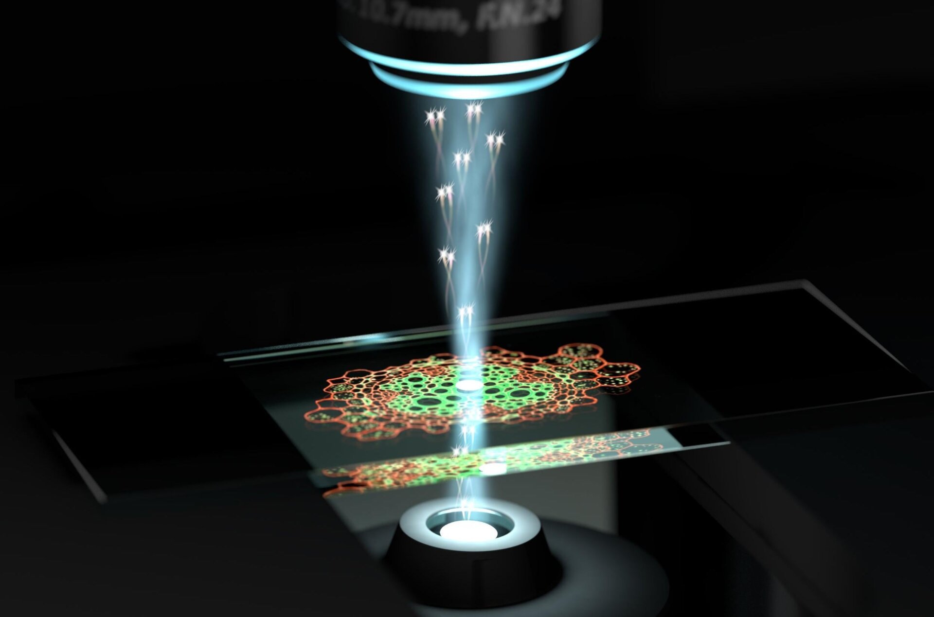
Quantum entanglement, a principle once confined to theoretical physics, is now revolutionizing microscopy. A team of researchers from Germany and Australia has developed a quantum optical microscope capable of imaging nanoscale biological structures with unprecedented resolution, all while keeping the living cells intact. This groundbreaking technique, which utilizes laser light millions of times brighter than the sun, holds immense potential for advancements in biomedical research and navigation technologies.
The core of this innovation lies in the use of quantum entangled photons. Entanglement creates an interdependence between two particles; measuring the properties of one instantly reveals the properties of its partner. This interconnectedness allows the microscope’s sensor to achieve higher resolution without inflicting the typical damage associated with high-intensity light sources. The team’s findings, published in Science, demonstrate that quantum correlations enable enhanced contrast and clarity, surpassing the limitations of conventional microscopy imposed by photodamage.
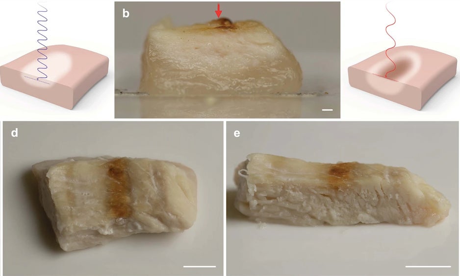
Traditional microscopes, while instrumental in advancing our understanding of biology, face limitations when observing delicate structures. The intense laser light required for high-resolution imaging can degrade or even destroy the target, much like a magnifying glass scorching ants under the sun. This photodamage restricts the level of detail achievable with conventional methods.
This new quantum microscope overcomes this hurdle. By employing entangled photon pairs, the researchers were able to image a yeast cell with remarkable clarity. The detection of one photon provides information about the arrival time of its entangled partner, effectively enhancing the resolution without increasing the laser’s intensity. The technique relies on Raman scattering, a method that analyzes the scattering of photons off molecules to determine their vibrational signature, effectively creating a chemical fingerprint.
The researchers successfully resolved the yeast cell’s cell wall, a nanometer-thick structure notoriously difficult to visualize with conventional Raman microscopes. This achievement highlights the quantum microscope’s ability to reveal intricate details previously obscured by photodamage.
This breakthrough has far-reaching implications. While Raman microscopy has long held promise for revolutionizing biological imaging, its limitations have hampered progress. The quantum approach offers a practical path forward, overcoming these obstacles and unlocking new possibilities for studying living cells at the nanoscale. This technology is still in its early stages, but the proof-of-principle demonstrated by this research paves the way for future advancements in biomedical imaging and beyond. From understanding fundamental biological processes to developing innovative diagnostic tools, the potential of quantum microscopy is vast.
The ability to observe the intricate machinery of life at such a fine scale opens up new avenues for scientific discovery. This quantum leap in microscopy promises to further unravel the complexities of the biological world, pushing the boundaries of our understanding and paving the way for groundbreaking advancements in various fields.
https://dx.doi.org/10.1038/s41586-021-03528-w
https://www.rp-photonics.com/laser_induced_damage.html
https://www.ibm.com/ibm/history/ibm100/us/en/icons/microscope/
https://www.sciencedirect.com/topics/medicine-and-dentistry/raman-scattering


