Corneal injuries, often treatable with conventional methods like corneal grafts, can sometimes be so severe that they deplete the eye’s supply of limbal epithelial stem cells. These stem cells are crucial for regenerating the cornea’s surface. Without them, the cornea remains damaged, and even a corneal graft won’t offer a lasting solution. However, promising new research offers a potential breakthrough for these challenging cases.
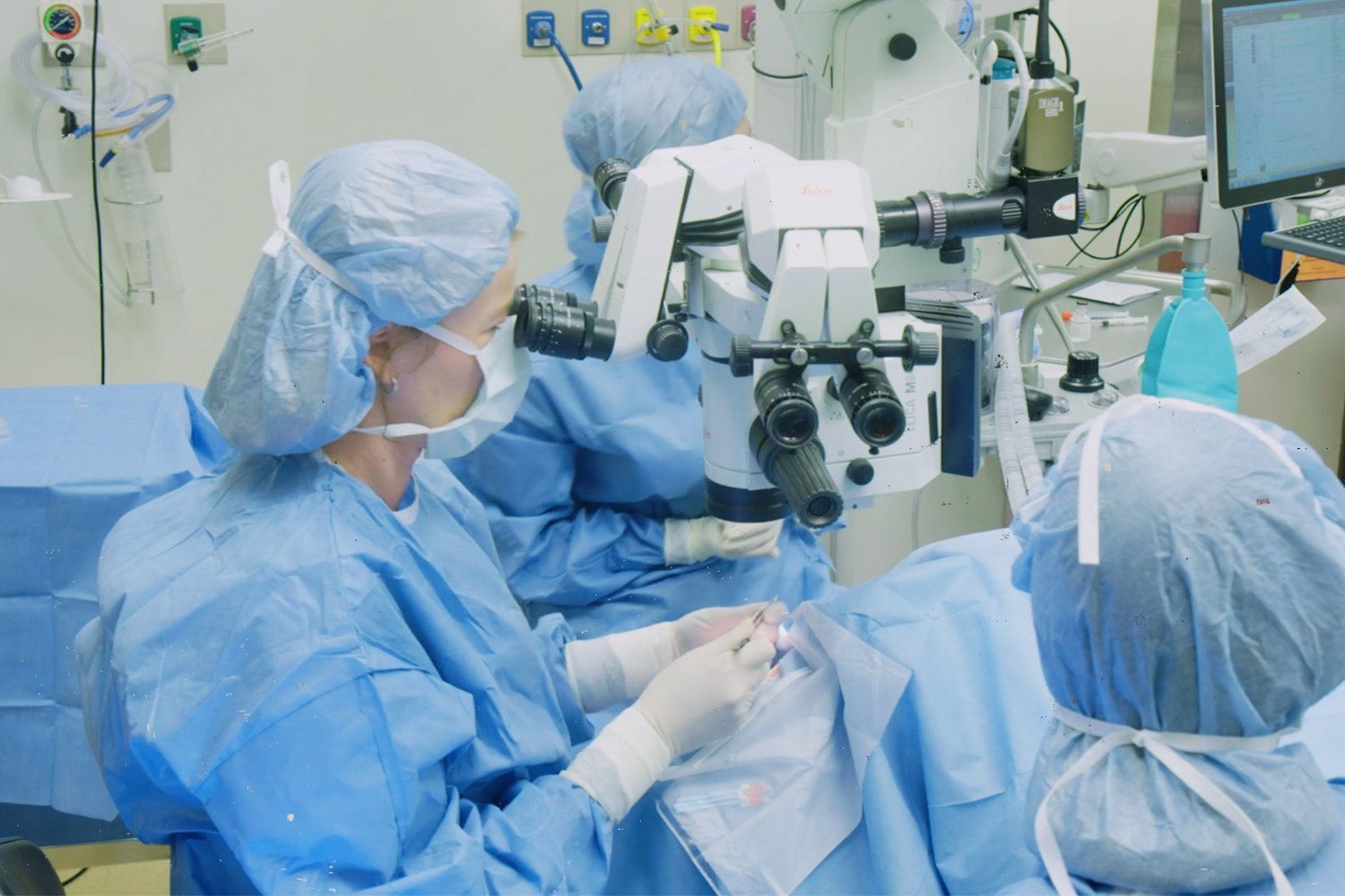
A Phase I/II clinical trial led by researchers at Mass Eye and Ear has shown encouraging results using cultivated autologous limbal epithelial cells (CALEC). This innovative therapy involves transplanting stem cells harvested from the patient’s healthy eye to the injured one. The study, involving 14 patients, demonstrated the potential of CALEC to safely restore the corneal surface and improve vision in individuals with previously untreatable injuries.
CALEC: A Novel Approach to Corneal Repair
The cornea, the transparent front layer of the eye, plays a vital role in protecting the eye and focusing light onto the retina for clear vision. Serious injuries or infections can scar the cornea, and while corneal grafts are often effective, they are insufficient when limbal stem cell deficiency occurs. This deficiency leaves the corneal surface permanently damaged, rendering traditional grafts ineffective. Lead study researcher Ula Jurkunas, associate director of the Cornea Service at Mass Eye and Ear, explains that limbal stem cell deficiency is a devastating condition characterized by a white cornea, vision loss, and significant pain. Until now, effective treatment options have been limited.
 1. First Calec Patient
Promising Results and Future Directions
The CALEC procedure involves carefully collecting and cultivating healthy stem cells from the patient’s uninjured cornea. These cells are then formed into a tissue graft and transplanted onto the damaged cornea. Previous research with four patients had indicated the safety and short-term efficacy of CALEC grafts. The latest study, published in Nature Communications, tracked 14 patients for up to 18 months post-procedure, revealing even more promising long-term results.
Significant Improvements in Corneal Health and Vision
The results of the study are compelling. Ninety-two percent of the patients showed at least partial improvement after a year and a half, with 77% achieving complete corneal surface restoration. Three patients received a second graft, with one achieving complete restoration afterward. Furthermore, all participants experienced some degree of visual acuity improvement. The procedure was well-tolerated, with no serious adverse events directly attributed to the CALEC transplant. One patient experienced a bacterial infection several months later, but this was linked to chronic contact lens use.
Jurkunas highlights the transformative impact of CALEC on patients’ lives, citing one patient who regained their quality of life after the procedure. While CALEC is still experimental and some patients may require an additional corneal graft for substantial vision improvement, this represents a significant advancement in treating corneal blindness using adult-derived stem cells.
Expanding Access and Refining the Technology
The research team is currently working on larger clinical trials across multiple eye centers. While the procedure isn’t yet widely available, they are also focusing on technological advancements, such as developing methods to cultivate and transplant stem cells from donors. This would expand access to the therapy for individuals with damage to both corneas. If further research continues to demonstrate the efficacy of CALEC, it could become a standard treatment for these previously irreversible conditions, offering renewed hope for patients suffering from severe corneal injuries.


