The European Synchrotron Radiation Facility (ESRF) in France has unveiled the “Extremely Brilliant Source,” the world’s brightest X-ray source. This groundbreaking technology, 10 trillion times more powerful than medical X-rays, is poised to revolutionize scientific research by enabling 3D imaging of matter at the atomic level. Scientists are already utilizing this powerful tool to study everything from the intricacies of the COVID-19 virus to the inner workings of fossils and batteries.
The Extremely Brilliant Source produces X-rays of unprecedented power, allowing scientists to create incredibly detailed 3D images. While a standard medical X-ray reveals the structure of bones and tissues, this advanced technology could theoretically visualize individual atoms within a blood cell. This immense power opens up endless possibilities for scientific exploration. ESRF Director General Francesco Sette highlights the potential for groundbreaking research on brain structure and function, paving the way for brain-like electronics.
Unveiling Microscopic Secrets
The applications of the Extremely Brilliant Source are vast and varied. Engineers can gain invaluable insights into innovative materials, advancing fields like aeronautics and nanoelectronics. Paleontologists can explore the intricate internal structures of fossils without damaging precious specimens. Crucially, researchers have already used the facility to image the lungs of COVID-19 victims, revealing previously unseen microscopic damage caused by the virus.
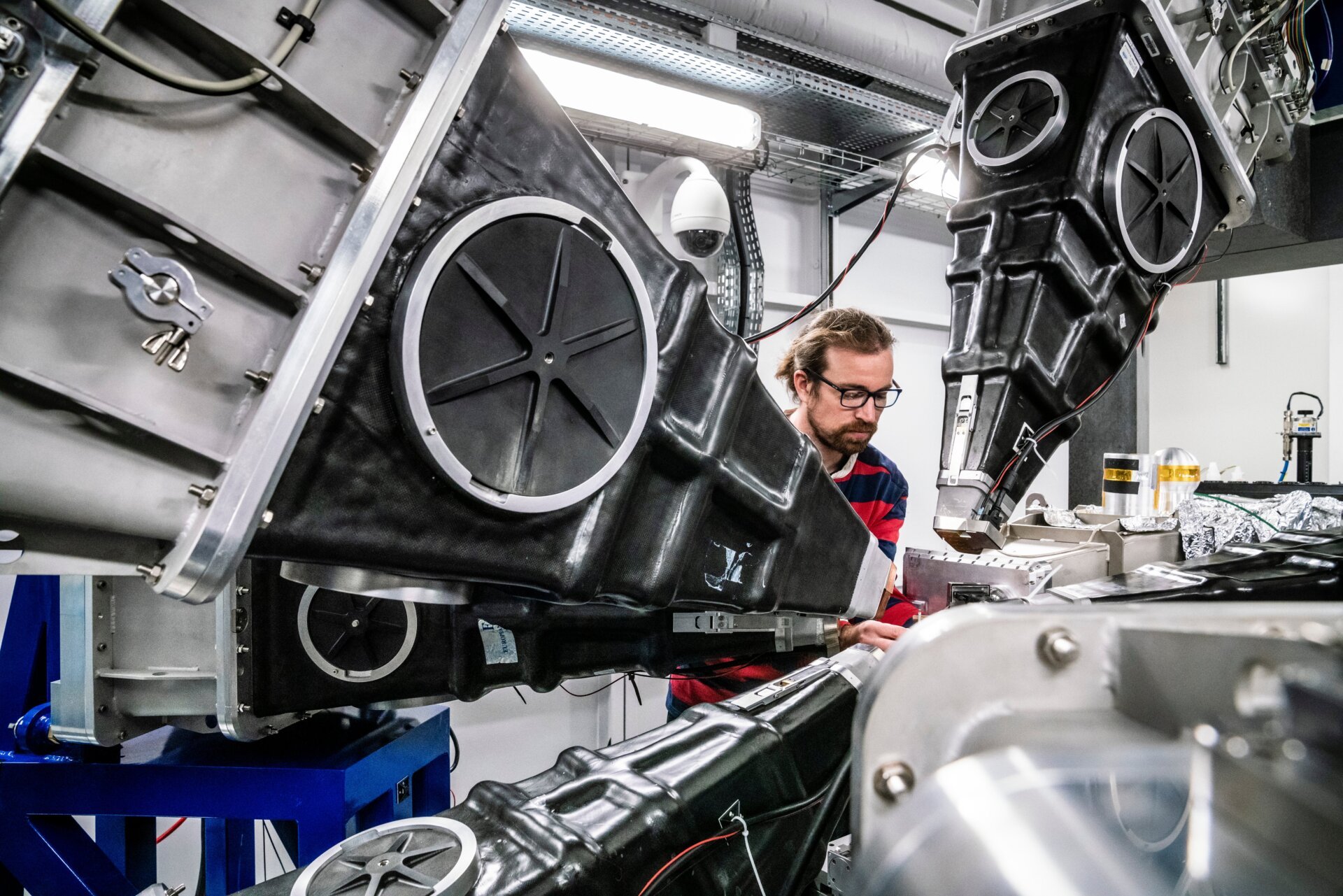 A researcher works at one of the European Synchrotron beamlines.A researcher operates equipment at a European Synchrotron beamline. Photo: S. Candé/ESRF
The Science of Synchrotrons
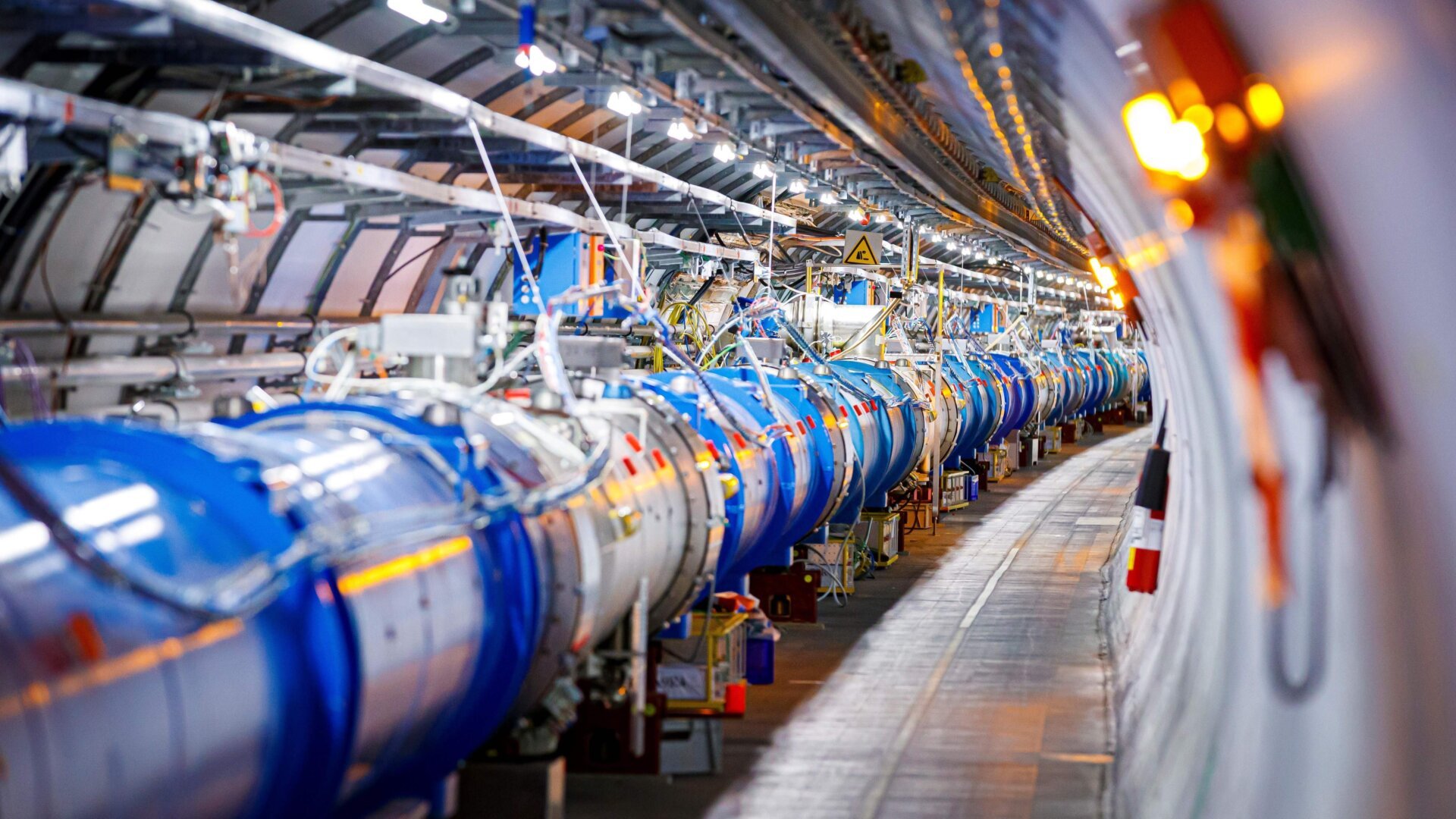
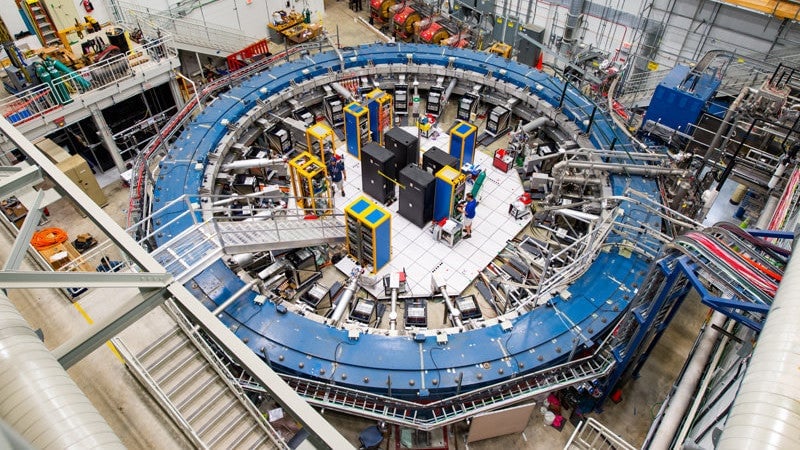
A synchrotron accelerates electrons using magnetic fields, generating high-energy X-ray light, also known as synchrotron light. Unlike colliders like the Large Hadron Collider, the particles in a synchrotron don’t collide. Instead, the X-rays emitted by the circulating electrons are directed into specialized laboratories called beamlines, where researchers use them for imaging. Synchrotron science has already led to remarkable discoveries, including visualizing the inside of a dinosaur egg and deciphering an ancient, volcano-damaged book. (https://www.newyorker.com/magazine/2015/11/16/the-invisible-library)
A Powerful Upgrade
The ESRF, operational since 1994, previously housed the world’s most powerful X-ray source. This latest upgrade increases its power by a factor of 100. The facility underwent a two-year shutdown for the transition, officially reopening in August 2020, ahead of schedule. Initial research results from the Extremely Brilliant Source are expected soon.
A New Era of X-Ray Imaging
The key to this upgrade lies in the redesigned lattice of 1,100 magnets that guide the electrons around the 844-meter ring. These magnets accelerate the electrons and provide directional “kicks,” generating the X-rays. As Sette explains, deviating the trajectory of a charged particle produces light—synchrotron light. This process adheres to the law of conservation of energy: the electrons lose energy when changing direction, emitted as light. The new magnet design allows continuous bending and refocusing of the electron beam, maximizing high-energy X-ray production without requiring a larger facility.
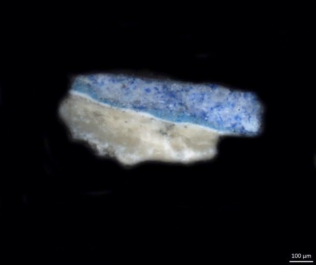 A sample of paint from Leonardo da Vinci’s “The Last Supper.” This sample is currently being analyzed at the ESRF.A paint sample from da Vinci’s “The Last Supper” undergoes analysis at the ESRF. Image: Courtesy Dipartimento di Fisica, Politecnico di Milano
Transforming Histology
Synchrotron science is poised to transform histology, the microscopic study of tissues. Traditional histology involves slicing and staining tissues, while synchrotron imaging allows for non-invasive, high-resolution 3D scans of whole samples, providing a much more comprehensive view of tissue anatomy. This “3D nano-histology” represents a revolutionary advancement in the medical field.
Accelerating Research
Victor Gonzalez, a postdoctoral researcher at the Rijksmuseum, uses synchrotron imaging to study historical paint samples, recently uncovering insights into Rembrandt’s techniques (https://www.sciencedaily.com/releases/2019/01/190114100152.htm). He emphasizes the upgrade’s significance, enabling faster analysis and new opportunities to understand the chemical processes in historical paints.
The Future of Scientific Discovery
With the Extremely Brilliant Source operational, scientists worldwide can apply for beamline time. While access is competitive, the upgrade significantly reduces experiment durations, from weeks to days, and from days to minutes. This powerful tool promises a wealth of exciting new scientific discoveries in the near future.


